Multiphoton Microscopy Imaging Technology No. 55: Self-localized Ultrafast Pencil Beams for Volumetric Multiphoton Imaging
 11/09 2026
 571
In multiphoton microscopy imaging, rapid acquisition of three-dimensional volumetric information requires a sufficiently long depth of focus. Bessel beams are currently the most commonly used long-depth-of-focus beams; they can maintain tight transverse confinement over a large axial range, but their pronounced side lobes and sensitivity to aberrations limit their use in complex biological tissues.
Another related physical phenomenon is that when ultrafast pulses are coupled into a multimode fiber (MMF) near the critical self-focusing power, the optical field spontaneously collapses into a pencil beam. This phenomenon has been predicted theoretically, but there have been few reports of its application to imaging. Recently, Honghao Cao and colleagues at MIT observed a self-localized ultrafast pencil beam in a standard multimode fiber for the first time and applied it to volumetric multiphoton imaging. The beam combines a long depth of focus, low noise, and high aberration tolerance, offering an alternative to Bessel beams for deep-tissue three-dimensional imaging [1].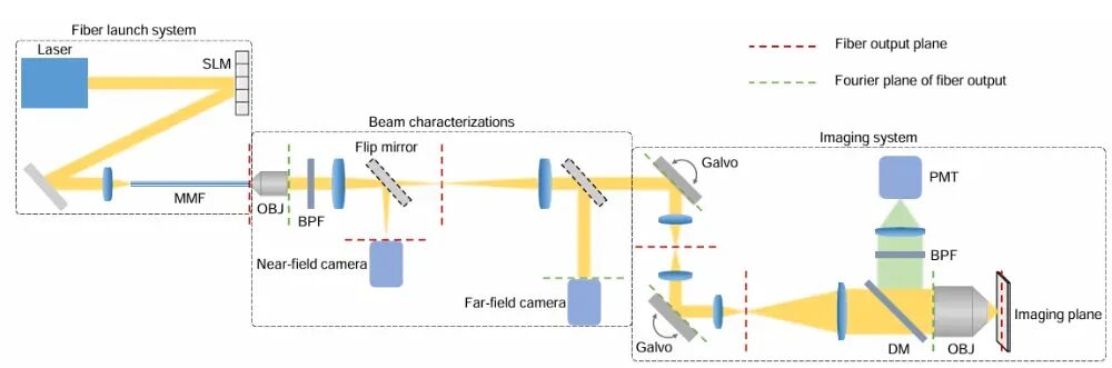
Figure 1 Schematic diagram of the experimental setup [1]
The experimental system consists of three parts: fiber injection, beam characterization, and two-photon imaging, as shown in Figure 1. A 1030 nm, 219 fs Gaussian pulse is precisely aligned via a spatial light modulator (SLM) and injected into a 50 μm core-diameter step-index multimode fiber, with a fiber output efficiency of approximately 8%. The emitted beam is scanned by a pair of galvanometer mirrors and then enters an objective with a numerical aperture of 1.05, and the two-photon fluorescence signal is collected by a photomultiplier tube.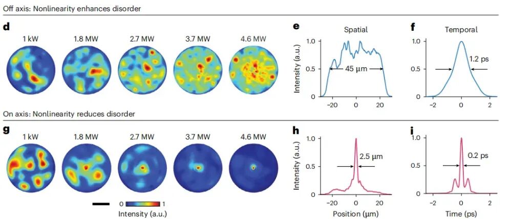
Figure 2 Off-axis injection nonlinearly enhances speckle disorder, whereas on-axis injection causes the speckle to contract into a pencil beam [1]
Under off-axis injection, as the peak power increases from 1 kW to 4.6 MW, the disorder of the output speckle pattern increases through nonlinear enhancement, and both spatial and temporal characteristics degrade. After aligning the incident light with the fiber axis, the speckle gradually contracts as the power increases, converging at 4.6 MW into a pencil beam with a transverse beam waist of 2.5 μm and a pulse width of 0.2 ps, as shown in Figure 2. This result indicates that at the critical self-focusing power, the optical field achieves ordering simultaneously in both spatial and temporal dimensions.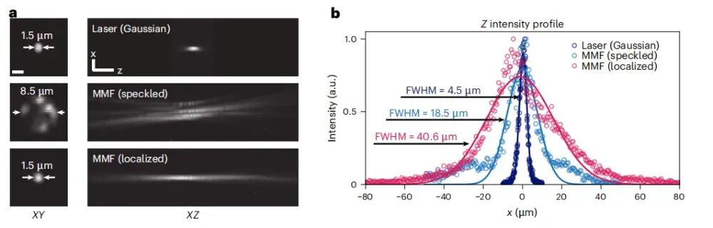
Figure 3 Comparison of the three-dimensional PSF of the self-localized beam axis and a Gaussian beam [1]
Three-dimensional point spread function (PSF) measurements (Figure 3) show that the self-localized beam has a transverse full width at half maximum (FWHM) of 1.5 μm, comparable to that of a Gaussian beam; its axial FWHM is approximately 40.6 μm, about ten times that of a Gaussian beam (4.5 μm) and also larger than the axial range of ordinary multimode fiber speckle (18.5 μm), with a more symmetric distribution and no obvious side lobes. This characteristic meets the requirements of volumetric imaging for focal spot shape, namely extending the axial coverage range while maintaining transverse resolution.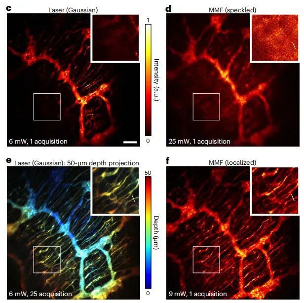 Figure 4 Two-photon imaging of the myenteric plexus of the small intestine driven by the self-localized beam and a Gaussian beam [1]
Two-photon imaging was performed using the myenteric plexus of the small intestine of adult mice as a sample. A Gaussian beam requires stitching 25 frames to obtain a clear projection within a 50 μm depth range, and the signal-to-noise ratio of a single frame is limited; the self-localized beam can obtain low-noise three-dimensional structural information in a single frame. The acquisition time for three-dimensional data was shortened from the 25-minute scale to the 1-minute scale, demonstrating the superiority of this technology in clinical and high-throughput screening scenarios.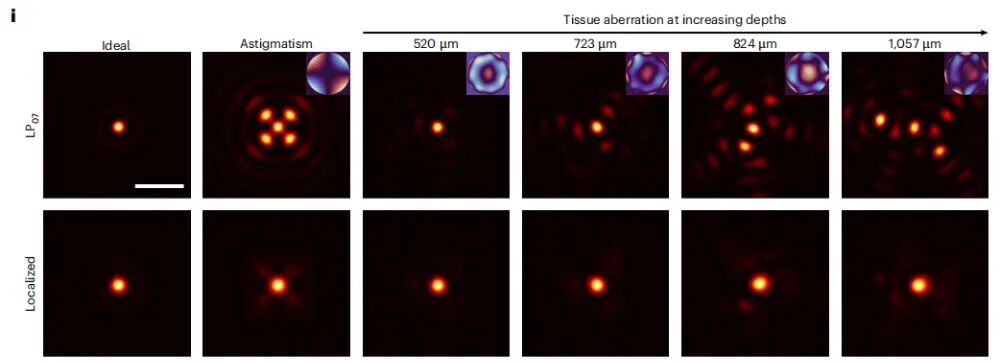 Figure 5 LP0n higher-order modes split and distort under depth aberrations, whereas the self-localized combined beam maintains a central bright spot [1]
The authors modeled the self-localized beam as an incoherent superposition of a set of higher-order LP0n modes (n = 6–10) and numerically simulated the influence of tissue aberrations (including astigmatism) increasing with depth. The results show that when the depth increases from 520 μm to 1057 μm, a single LP0n mode rapidly splits and distorts, whereas the self-localized combined beam always maintains a compact central bright spot, demonstrating robustness against aberrations in complex tissue.
At present, this technology still has certain limitations: it has only been validated at two wavelengths, 1030 nm and 1300 nm; the upper limit of input peak power is 1 μJ, and the fiber output efficiency is only 8% (about 80 nJ); the beam is relatively sensitive to incident angle and power; and the related theoretical analysis remains to be improved. Subsequent work plans to further optimize beam quality by increasing the repetition rate (currently 1 MHz) and through pulse compression.
The significance of this work lies in converting the speckle problem caused by nonlinear effects in multimode fibers into an exploitable physical resource: only a segment of 50 μm step-index multimode fiber and a spatial light modulator are needed to convert a conventional femtosecond laser into a light source suitable for volumetric imaging. The system is simple in structure, low in cost, and stable in performance, providing a feasible engineering solution for clinical and point-of-care applications of multiphoton microscopy.
References [1] Cao H, et al. Self-localized ultrafast pencil beam for volumetric multiphoton imaging. Nature Methods, 2026. DOI: 10.1038/s41592-026-03067-0.
-
![]()
Thailand Begins to Settle Old Scores with Chinese Automakers
-
![]()
Why Did It Take smart Seven Years to Perfect a Small Car Priced at 99,900 Yuan?
-
![]()
Tesla's All-Electric Pickup Cybertruck Is About to Be Discontinued: 70,800 Units Sold in 33 Months, Production Line to Be Freed Up for Cybercab in Mid-2027
-
![]()
OpenAI Account Bans Are Just the Tip of the Iceberg: The "Reckoning" of AI Compute Is Unfolding Simultaneously in Washington and Wall Street
-
![]()
Multiphoton Microscopy Imaging Technology No. 55: Self-localized Ultrafast Pencil Beams for Volumetric Multiphoton Imaging
-
![]()
China and the US have already settled their AI competition—and it's not even close: Multiple US states move to halt data center construction
-
![]()
Huguang Shares: Revenue Down 8.05%, Profit Plummets 80.25% Amid Wave of New Designations, Gross Margin Falls 3.74 Points
-
![]()
BYD Executive Responds to Maserati Acquisition Rumors: BYD's Cars Are the Fastest in the World